Union Budget 2024: 1 फरवरी 2024 को वित्तमंत्री निर्मला सीतारमण छठी बार बजट पेश करेंगी। निर्मला सीतारमण दूसरे कार्यकाल का आखिरी बजट पेश करेंगी। इसके बाद देशभर में लोकसभा चुनाव होंगे। वित्तमंत्री सरकार को वोट बैंक भरने के लिए कई घोषणाएं कर सकती हैं। विशेषकर सरकारी कर्मचारियों के लिए। सरकार चुनाव से पहले लोगों को लाभान्वित करने के लक्ष्य में हो सकती है। यह एक महत्वपूर्ण समय है जब सरकार अपने कार्यों को सार्थकता देने का प्रयास करेगी।
Union Budget 2024: वेतन में आएगा बंपर उछाल
निर्मला सीतारमण 1 फरवरी 2024 को छठी बार बजट पेश करेंगी। वह मोदी सरकार के आखिरी कार्यकाल का आखिरी बजट पेश करेंगी। इसके बाद देशभर में लोकसभा चुनाव होंगे। सरकार वोट बैंक को भुनाने के लिए कई घोषणाएं कर सकती है। सरकारी कर्मचारियों के लिए महंगाई भत्ता बढ़ाने का विचार है।
बजट से पहले कर्मचारियों का महंगाई भत्ता 50% तक बढ़ा सकती है। सरकार साल में दो बार महंगाई भत्ता बढ़ाती है। इस बार उम्मीद है कि बजट में 50% महंगाई भत्ते की घोषणा हो। निर्मला सीतारमण की दूसरे कार्यकाल की आखिरी बजट होगी। सरकार लोकसभा चुनाव से पहले कर्मचारियों की भलाइयों का ध्यान रख सकती है।
चुनावों से पहले कर्मचारियों के लिए होंगे बड़े ऐलान
- सरकार के वोट बैंक के कर्मचारी नए साल में बढ़ती सैलरी की उम्मीद से भरे हैं।
- 2024 में वित्त मंत्री निर्मला सीतारमण अंतरिम बजट पेश करेंगी।
- चुनावी साल के कारण सरकार संभावित डीए बढ़ाने का विचार कर रही है।
- उम्मीद है कि सरकार चुनावों से पहले कर्मचारियों की सैलरी में इजाफा करेगी।
- सरकार डीए बढ़ाने का ऐलान 2024 में चुनावों से पहले कर सकती है।
- ताकि सरकारी कर्मचारियों को चुनावों से पहले लुभा सके।
- ऐसा होने पर कर्मचारियों को सैलरी और भत्तों में वृद्धि हो सकती है।
- सरकार का इस पर ऐलान चुनावी माहौल में लोकप्रिय हो सकता है।
- कर्मचारियों को इस कदम से जुड़ी ऊर्जा महसूस होगी।
- सरकारी कर्मचारियों के लिए इस वर्ष का आरंभ उत्साहपूर्ण हो सकता है।
महंगाई भत्ते का नियम
- साल 2016 में 7वां वेतन आयोग लागू होने पर, महंगाई भत्ता शून्य हो गया।
- नियम के अनुसार, जब भत्ता 50% तक पहुंचेगा, तो वह शून्य किया जाएगा।
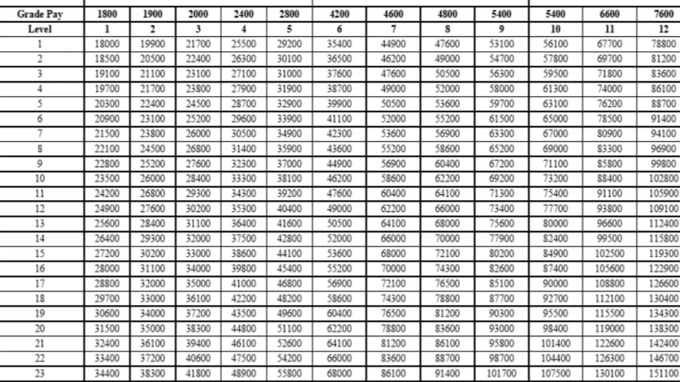
- कर्मचारियों को मिलने वाला पैसा 50% बेसिक सैलरी में जोड़ा जाएगा।
- किसी की बेसिक सैलरी 18000 रुपये होने पर, उसे 9000 रुपये महंगाई भत्ता मिलेगा।
- लेकिन, डीए 50% होने पर यह बेसिक से जुड़ जाएगा और भत्ता जीरो होगा।
सरकार ने निर्णय से कर्मचारियों को संबोधित किया। महंगाई भत्ता बचाने के लिए कर्मचारियों को बेहतर सुविधाएं मिलेंगी। यह नियम भत्ते की प्रणाली में सुधार करने का प्रयास है। कर्मचारियों की आय में सुधार होने की उम्मीद है। वेतन वृद्धि से महंगाई को संतुलित करने का प्रयास किया गया है।
| Whatsapp Channel | Join |
| Telegram Channel | Click Here |
| Homepage | Click Here |